⚖️ UK Legal Notice: This guide is written strictly for educational, taxonomic, and microscopy research purposes. Psilocybe cubensis spores do not contain psilocybin or psilocin and are legal to purchase in the UK for microscopy research. Growing fruiting mushrooms from spores is illegal under the Misuse of Drugs Act 1971. Always comply with UK law. See our 2026 UK Legal Guide for full details.
⚡ Quick Answer
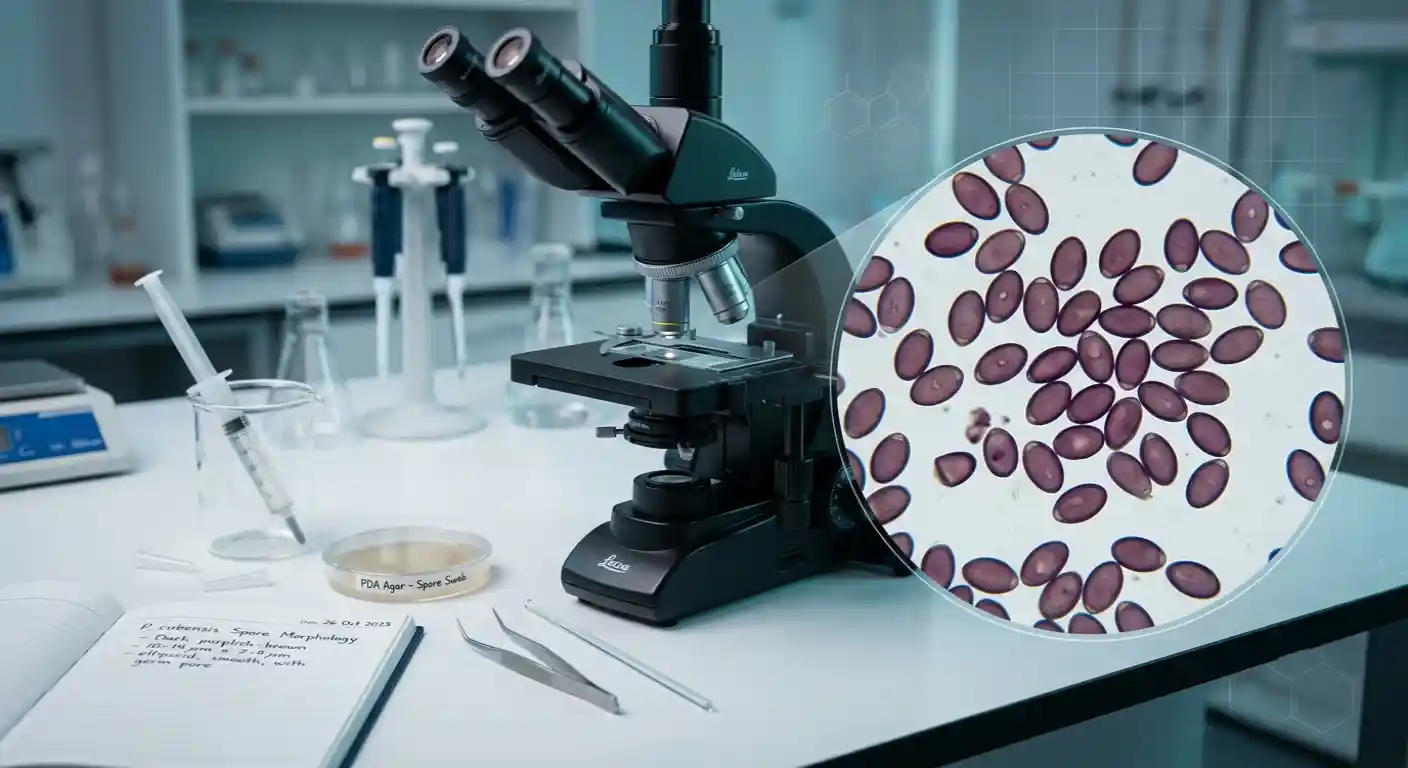
Under a microscope, Psilocybe cubensis spores appear as small, brown to purple-brown, oval shapes (some slightly angular face-on) with a smooth, thick wall and a visible thin-walled "germ pore" at one tip. They measure about 11.5–17 µm long and 6–11.5 µm wide (Singer & Smith, 1958; Batista et al., 2025), whichever strain they come from, which is about one-fifth the width of a human hair. They're clearly visible at 400x magnification, but you'll need 1000x oil immersion to make out fine detail like wall thickness and the germ pore. This guide breaks down exactly what to expect at each stage of magnification, and what each visual feature actually means.
Psilocybe cubensis spore morphology: oval, brown to purple-brown, smooth thick wall, visible germ pore
11.5–17 µmTypical spore length across cubensis strains
Brown to Purple-BrownStandard spore pigmentation
400xMinimum magnification to see spores clearly
1000xMagnification for fine detail (oil immersion)
Jump to a section
1. Basic Appearance
2. Size and Measurements
3. Colour and Pigmentation
4. Shape, Wall and Germ Pore
5. What You See at Each Magnification
6. How to Observe Spores Properly
FAQ1. What Do Cubensis Spores Look Like, Overall?
At first glance under a microscope, Psilocybe cubensis spores look like scattered dark oval grains — a bit like tiny coffee beans, though far more uniform in shape. They're not transparent; the dark pigmentation is one of the most useful features for spotting them quickly, even at low magnification. Every named cubensis strain — Golden Teacher, B+, Penis Envy, and the rest — shares this same basic appearance, because they're all the same species. Strains can't be told apart by their spores: they are cultivated varieties of one species, and their spores overlap in size, shape and colour.
ℹ️ Key Identification Fact — Dark pigmentation, an oval (subellipsoid) shape, and a visible germ pore are the three features that most reliably identify Psilocybe spores under a microscope, distinguishing them from look-alike genera.
2. How Big Are Cubensis Spores?
Psilocybe cubensis spores typically measure about 11.5–17 µm long and 6–11.5 µm wide (Singer & Smith, 1958; Batista et al., 2025), whichever strain they come from. A micrometre is one millionth of a metre: for scale, a human hair is roughly 70µm wide, so a cubensis spore is about one-fifth the width of a single hair.Golden Teacher
11.5–17 µm
The normal P. cubensis range, the same for every strain.
B+
11.5–17 µm
Same range as Golden Teacher.
Penis Envy
11.5–17 µm
Same size range as Golden Teacher; no published difference in wall thickness.
3. What Colour Are Cubensis Spores?
Under the microscope, cubensis spores look brown to purple-brown; the exact shade depends on lighting, mounting liquid and how thickly they're spread. This dark pigmentation gives them strong contrast against the pale background of a glass slide under standard brightfield illumination — you don't need special staining or lighting techniques to see them clearly. The spores themselves are pigmented, which is why they show up without staining. A single spore looks lighter than a spore print: one spore lets more light through, while a print or thick deposit (thousands of spores piled together) looks dark purple-brown to almost black.
✅ Colour Check — Colour alone isn't a reliable test. Genuine cubensis spores are brown to purple-brown, oval and smooth, with a germ pore at one end. Bright yellow, white or highly irregular objects are more likely contamination (mould spores) than cubensis spores.
4. Shape, Wall, and Germ Pore
Beyond colour and size, three structural features define what you're looking at.
🥚
Shape — Subellipsoid
Roughly oval but slightly flattened, so it looks a little different side-on and face-on, and some look slightly angular face-on. Picture a rugby-ball shape with one side gently pressed flat. Most spores in a healthy sample look broadly similar.
🧱
Wall — Smooth
The outer wall is smooth with no warts, spines, or surface ornamentation, typically 0.5–1.0µm thick. Only clearly visible at 1000x oil immersion.
🔲
Germ Pore — Visible Window
A small, thin-walled area at one tip of the spore. It looks like a slightly lighter "window" set into the darker spore body, one of the clearest features separating Psilocybe from look-alike genera.
📏
Consistency
In a clean sample, spores look broadly similar in shape and colour, with a little natural variation from spore to spore. Objects that look very different in shape or structure may be contamination, debris or a degraded sample.5. What You Actually See at Each Magnification
What's visible changes significantly as you step up magnification. Here's a realistic breakdown of what to expect.| Magnification | What You'll See | Best For |
|---|---|---|
| 100x | General overview, spore clusters as dark specks | Finding a good area of the slide to focus on |
| 400x | Clear individual spore shape, colour, and general distribution | Standard observation, size estimation, taking measurements |
| 1000x (oil immersion) | Wall thickness, germ pore detail, fine surface texture | Detailed structural analysis, strain comparison work |
6. How to Observe Spores Properly
Getting a clear view depends as much on slide preparation as on your microscope.1
Clean your slide — Wipe with isopropyl alcohol and lens tissue, and let it dry fully. Dust and fingerprints create artefacts that can be mistaken for contamination.
2
Agitate the spore syringe — Roll it between your palms for 10–20 seconds to redistribute settled spores evenly through the liquid.
3
Add one small drop — Around 0.05ml onto the centre of the slide. Too much liquid causes the coverslip to float and the spores to drift out of focus.
4
Apply the coverslip at an angle — Let one edge touch the liquid first, then lower it flat to push air out and avoid bubbles.
5
Start at 100x, then move to 400x — Get an overview first, then switch up to your main working magnification to observe shape and colour.
6
Finish at 1000x oil immersion — Add a small drop of Type B immersion oil on the coverslip for the 100x oil objective to reveal wall and germ pore detail.
⚠️ One Thing to Remember — Always cap the needle immediately after drawing a drop. Leaving the syringe open to air risks contamination that will affect every future slide you prepare from it.
Ready to Start Observing?
Tripping Store's spore syringes are prepared under sterile conditions in the UK, with verified genetics and consistent, well-documented morphology — giving you a reliable starting point for microscopy research.Frequently Asked Questions
QWhat do Psilocybe cubensis spores look like under a microscope?
They appear as small, brown to purple-brown, oval shapes (some slightly angular face-on) with a smooth, thick wall and a visible germ pore at one tip. They measure about 11.5–17 µm long and 6–11.5 µm wide and are clearly visible at 400x magnification.
QWhat colour are cubensis spores?
Under the microscope, single spores look brown to purple-brown, depending on lighting and mounting liquid. A spore print looks darker, dark purple-brown to almost black, because thousands of spores are piled together.
QHow big are Psilocybe cubensis spores?
Psilocybe cubensis spores typically measure about 11.5–17 µm long and 6–11.5 µm wide (Singer & Smith, 1958; Batista et al., 2025), whichever strain they come from. A micrometre is one millionth of a metre: for scale, a human hair is roughly 70µm wide, so a cubensis spore is about one-fifth the width of a single hair.
QWhat magnification do I need to see spore detail?
You can see general shape and colour at 400x. For fine detail like wall thickness and the germ pore, you'll need 1000x magnification with oil immersion.
QWhat is the germ pore and why does it matter?
The germ pore is a small, thin-walled area at the tip of the spore. It's a key identification feature present in Psilocybe spores and is one of the clearest ways to distinguish them from look-alike genera at high magnification.
QCan spore appearance tell me which strain I have?
No. All cubensis strains share the same species-level appearance, and their spores look alike under the microscope. Differences in shade usually come from lighting, focus, mounting liquid or camera settings. Reliable strain identification needs documented provenance.
QHow do I know if what I'm seeing is contamination rather than spores?
Colour alone isn't a reliable test. Genuine cubensis spores are brown to purple-brown, oval and smooth, with a germ pore at one end. Bright yellow, white or highly irregular objects on your slide are more likely contamination such as mould.
QIs it legal to look at Psilocybe cubensis spores in the UK?
Yes. Psilocybe cubensis spores do not contain psilocybin or psilocin, so they are legal to buy, own, and study for microscopy and taxonomic research in the UK. Cultivating fruiting mushrooms from spores is not legal.